Description
Mitochondrial markers are structural or functional properties that are specific for mitochondria. In isolated mitochondria, protein can be determined as a measure of amount of mitochondria or mt-concentration [mg mt-protein/ml]. However, protein cannot be used as a mt-biomarker in other mitochondrial preparations. A structural mt-marker is the area of the inner mt-membrane or mt-volume determined stereologically, which has its limitations due to different states of swelling. If mt-area is determined by electron microscopy, the statistical challenge has to be met to convert area into a volume. When fluorescent dyes are used as mt-marker, distinction is necessary between mt-membrane potential dependent and independent dyes. mtDNA or cardiolipin content may be considered as a mt-marker. Determination of mitochondrial marker enzymes may be molecular (amount of protein) or functional (enzyme activities). Respiratory capacity in a defined respiratory state of a mt-preparation can be considered as a functional mt-marker, in which case respiration is expressed as flux control ratios. » MiPNet article
Abbreviation: mt-marker
Reference: Gnaiger 2014 MitoPathways
MitoPedia methods:
Respirometry
Mitochondrial markers and expression of mitochondrial respiration
| Gnaiger E (2014) Mitochondrial markers and expression of mitochondrial respiration. Mitochondr Physiol Network 2014-07-26. |
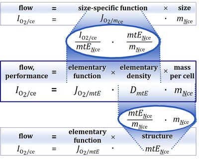
Abstract: Respiratory performance capacity of an organism, tissue or cell may change due to a change in size, concentration of functional elements (biomarker density) or element function (mt-specific function).
• O2k-Network Lab: AT Innsbruck Gnaiger E
Labels:
HRR: Theory
Biomarkers
Markers for size are volume, mass, area. If mass is not expressed as total mass but as protein mass, then a physical marker of size is replaced by a biochemical marker. Determination of the size of a system under investigation always requires definition of the system. The system (subject) whose phenotype is studied may be an organism, a tissue or a cell. The biochemical marker total protein can be replaced by a specific protein, e.g. by a marker enzyme.[1] A biomarker can be considered as a functional element. Expressing performance (IO2, oxygen flow per biological system) per size yields specific performance (JO2, oxygen flux = flow per system size) in an unstructured analysis.[2] A biomarker introduces a structural (and functional) element into the analysis. Therefore, expressing performance (IO2) per functional element (biomarker) yields marker-specific performance (Jmt,O2, mt-specific oxygen flux = flow per mt-marker) in a structured analysis.[3]
When replacing an enzyme activity by the respiratory activity in a defined respiratory state as a biomarker, then flow per mt-marker becomes a flux control ratio (FCR).[4],[5] Expressing structure by functional markers is without problem as long as the structure in question does not undergo any functional (qualitative) changes.
References
- ↑ Renner K, Amberger A, Konwalinka G, Kofler R, Gnaiger E (2003) Changes of mitochondrial respiration, mitochondrial content and cell size after induction of apoptosis in leukemia cells. Biochim Biophys Acta 1642: 115-23. »PMID: 12972300
- ↑ Gnaiger E (1993) Nonequilibrium thermodynamics of energy transformations. Pure Appl Chem 65: 1983-2002. »Open Access
- ↑ Gnaiger E (2014) Mitochondrial pathways and respiratory control. An introduction to OXPHOS analysis. 4th ed. Mitochondr Physiol Network 19.12. OROBOROS MiPNet Publications, Innsbruck: 64 pp. »Open Access
- ↑ Gnaiger E (2009) Capacity of oxidative phosphorylation in human skeletal muscle. New perspectives of mitochondrial physiology. Int J Biochem Cell Biol 41: 1837-45. »PMID: 19467914
- ↑ Pesta D, Hoppel F, Macek C, Messner H, Faulhaber M, Kobel C, Parson W, Burtscher M, Schocke M, Gnaiger E (2011) Similar qualitative and quantitative changes of mitochondrial respiration following strength and endurance training in normoxia and hypoxia in sedentary humans. Am J Physiol Regul Integr Comp Physiol 301: R1078–87. »Open Access


